Cell Division (Mitosis & Meiosis)
- One diploid cell into two identical daughtersnot yet tested
- Halving and shuffling the genome for gametesnot yet tested
- G1-S-G2-M checkpoints that fail in cancernot yet tested
- Eggs, stem cells, and the building of an embryonot yet tested
In 1879, the German anatomist Walther Flemming — using newly available aniline dyes that selectively stained the cell nucleus — watched a salamander cell divide under his microscope and produced the first detailed drawings of what we now call mitosis. The chromosomes (a name Wilhelm von Waldeyer-Hartz gave them in 1888 — coloured bodies) condensed, lined up, split, and migrated to two daughter nuclei. Over the following decades, the parallel process for gametes — meiosis — was worked out: a two-stage division that halves the chromosome number, producing eggs and sperm with one copy of each chromosome rather than two. Sexual reproduction required this halving, because fertilization would otherwise double the genome every generation.
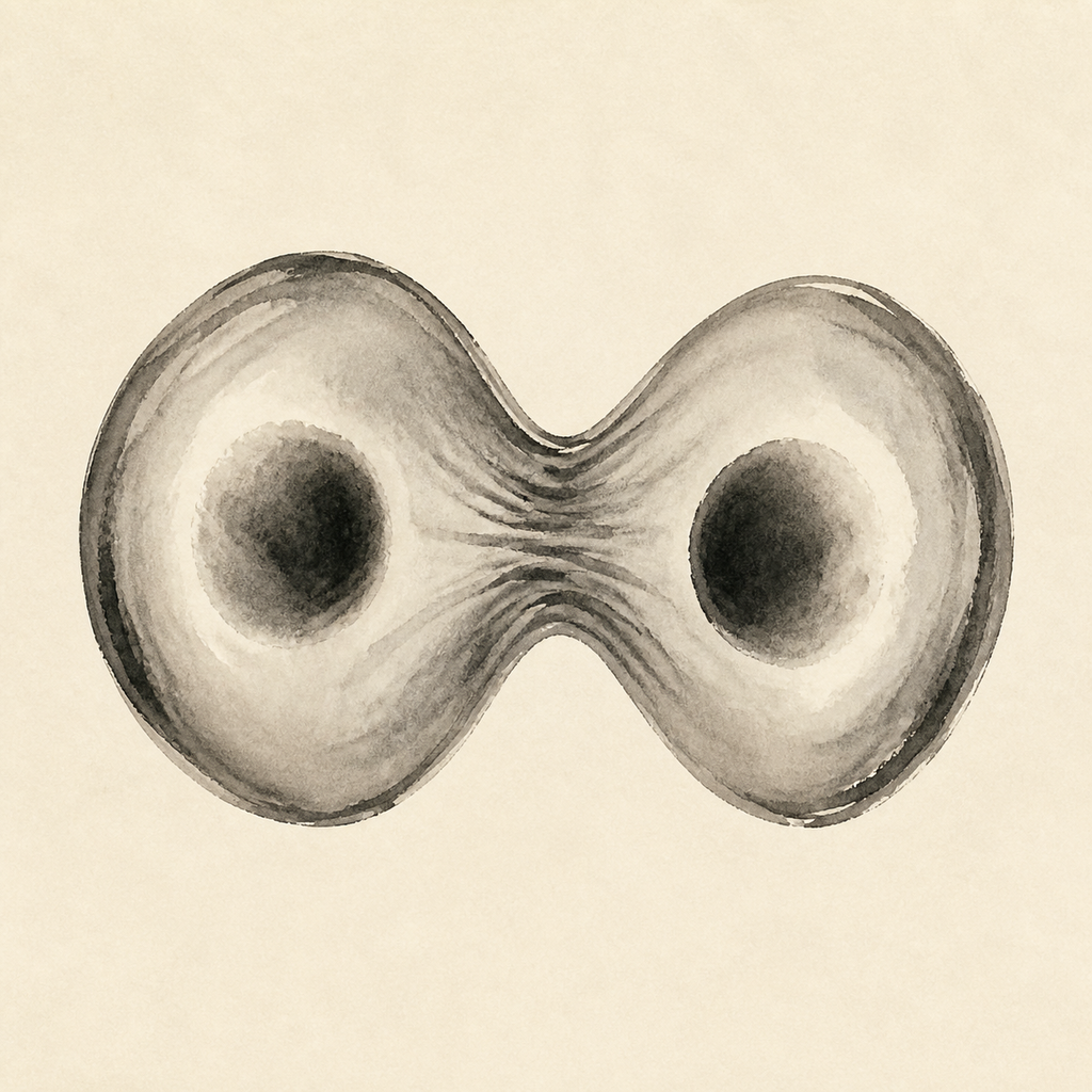
Mitosis is the somatic-cell version: one diploid cell, with two copies of each chromosome, becomes two diploid daughters carrying identical genomes. The chromosomes condense and the nuclear envelope dissolves; a mitotic spindle assembled from microtubules anchored at the centrosomes captures sister chromatids by their kinetochores from opposite poles; the chromatids align at the metaphase plate, separate cleanly, and migrate to opposite ends; new nuclear envelopes reform around each daughter set; and an actin-myosin contractile ring physically splits the cell in two. The whole sequence is embedded in a four-stage cell cycle — growth in G1, DNA replication in S, preparation in G2, division in M — and at every transition there are checkpoints run by cyclins, cyclin-dependent kinases, and guardian proteins like p53 and Rb that halt the cycle if DNA is damaged. Loss of these checkpoints is one of the defining features of cancer. Meiosis is the more interesting machine. Sexual reproduction requires gametes carrying half the parent's chromosome count, and meiosis solves the problem with two divisions on a single round of DNA replication. In meiosis I, the maternal and paternal copies of each chromosome pair up, exchange segments through crossing over — visible under the microscope as chiasmata — and separate into two cells. Meiosis II then separates the sister chromatids, producing four haploid daughter cells that are genetically distinct from one another. The two sources of diversity, crossing over and the independent assortment of homologous pairs, are what give natural selection its raw material. Errors in meiosis — non-disjunction, where chromosomes fail to separate cleanly — produce gametes with the wrong chromosome count, and the rate climbs with maternal age in particular.