Brain Anatomy
- Cerebrum, cerebellum, hemispheresnot yet tested
- Cortex and its four lobesnot yet tested
- Limbic system: seat of emotionnot yet tested
- Lesion landmarks: Phineas Gagenot yet tested
Weighing about 1.4 kilograms and packed with some 86 billion neurons, the human brain is not one organ but many, layered over each other in the order evolution added them. We know the map in remarkable detail, and we learned most of it the hard way — by watching what a person loses when a particular piece is damaged. A stroke that spares speech but erases the ability to form new memories; an injury that leaves intelligence intact but flattens all emotion: each deficit pins a function to a place. Read this way, the brain is less a uniform thinking-machine than a stack of specialized instruments, each added to solve a problem the previous one could not.
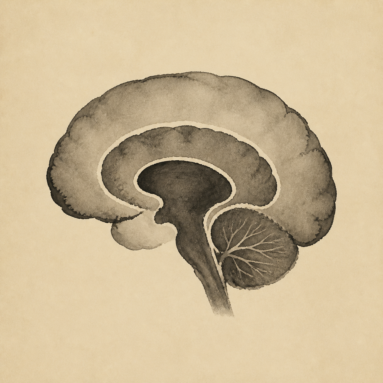
Read from the inside out, the brain climbs in evolutionary age. At the core sits the brainstem, the oldest part, running breathing and heartbeat and the sleep-wake cycle without ever consulting us — which is why an injury there is so often fatal. Tucked behind it, the cerebellum holds most of the brain's neurons in a fraction of its volume and quietly keeps movement smooth and timed. Above the brainstem, the thalamus works as a switchboard, routing nearly every sense up toward the higher brain, while the neighboring hypothalamus governs the body's thermostat, its hormones, its hungers. Wrapped around these lies the limbic system, the seat of memory and feeling — the hippocampus, without which no new experience can be laid down as memory, the lesson written into the amnesiac patient known only as H.M., and the amygdala, which stamps events with fear. Last and outermost comes the cerebral cortex, the crumpled sheet just millimeters thick that makes us recognizably human; its folds cram a newspaper-sized surface into the skull, and its four lobes split the labor — the frontal plans and speaks, the parietal locates the body in space, the temporal hears and understands, the occipital sees. The deep point is that this is not a chain of command but a collaboration of specialists: almost any region lies two or three synapses from any other, and even our highest thought is the old survival machinery pressed into new work.